Objective: To train and validate the use of a novel artificial intelligence-based
thermal imaging system as a screening tool to rule out malignancy in cutaneous
and subcutaneous masses in dogs.
Animals: Training study: 147 client-owned dogs with 233 masses. Validation
Study: 299 client-owned dogs with 525 masses. Cytology was non-diagnostic in
94 masses, resulting in 431 masses from 248 dogs with diagnostic samples.
Procedures: The prospective studies were conducted between June 2020 and
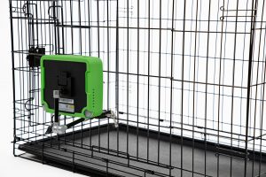
July 2022. During the scan, each mass and its adjacent healthy tissue was heated
by a high-power Light-Emitting Diode. The tissue temperature was recorded
by the device and consequently analyzed using a supervised machine learning
algorithm to determine whether the mass required further investigation. The first
study was performed to collect data to train the algorithm. The second study
validated the algorithm, as the real-time device predictions were compared to the
cytology and/or biopsy results.
Results: The results for the validation study were that the device correctly
classified 45 out of 53 malignant masses and 253 out of 378 benign masses
(sensitivity = 85% and specificity = 67%). The negative predictive value of the
system (i.e., percent of benign masses identified as benign) was 97%.
Clinical relevance: The results demonstrate that this novel system could be used
as a decision-support tool at the point of care, enabling clinicians to dierentiate
between benign lesions and those requiring further diagnostics.
Front. Vet. Sci., 29 September 2023
Sec. Oncology in Veterinary Medicine
Volume 10 – 2023 | https://doi.org/10.3389/fvets.2023.1164438


